Calcific tendinitis of the shoulder: when pain strikes without warning
Sudden, severe shoulder pain, sometimes in the middle of the night, with no injury before it - clinically different from ordinary tendon inflammation.
Calcific tendinitis of the shoulder causes sudden, severe pain, sometimes in the middle of the night, with no injury before it - completely different from ordinary tendon inflammation, and more common in women aged 30-50.
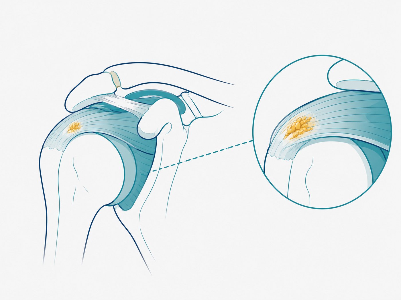
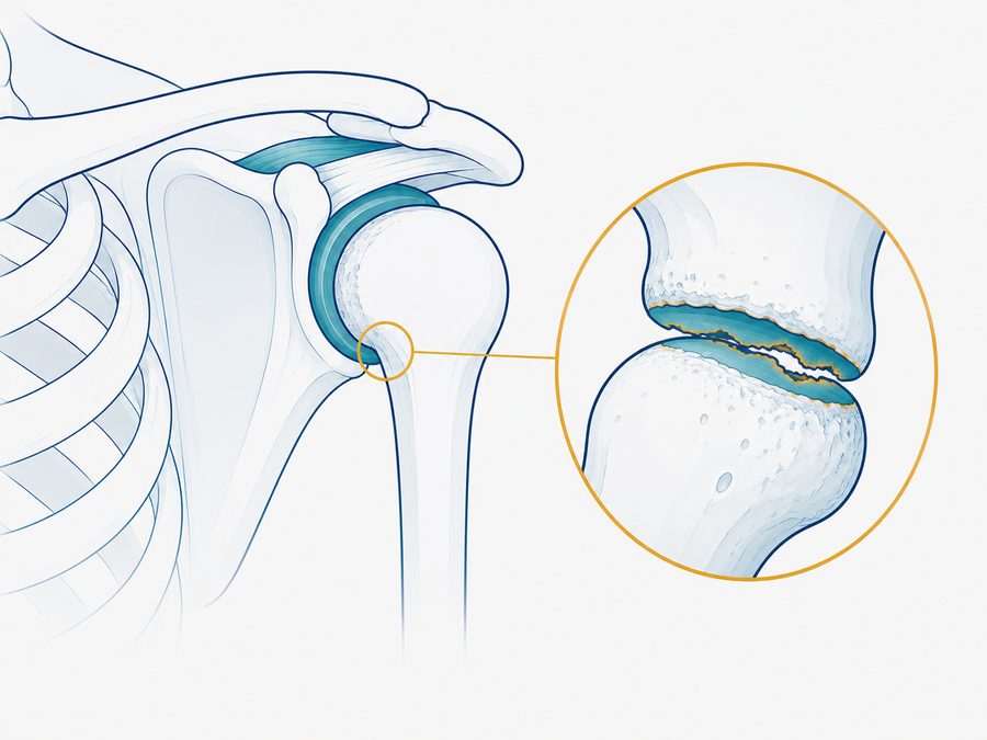
If you woke up in the middle of the night with sharp, unbearable shoulder pain and no fall or injury to explain it - this may be calcific tendinitis of the shoulder, a condition in which calcium deposits build up inside one of the shoulder tendons, usually the supraspinatus tendon.
This is an entirely different condition from “ordinary” tendon inflammation or a rotator cuff tear, and despite the dramatic pain it can cause, in most cases it responds well to conservative treatment.
How does it usually feel?
Symptoms can vary, but many describe:
- Sudden, severe shoulder pain, sometimes with no triggering event at all.
- Pain that gets much worse at night and disrupts sleep.
- Real difficulty with any shoulder movement, even the smallest.
- Sometimes also longer stretches of duller, milder pain.
- Tenderness in one spot at the front and top of the shoulder.
If the pain comes on extremely suddenly and is unusually intense, with no injury behind it - that is exactly the feature that sets active calcification apart from most other shoulder problems.
What causes calcific tendinitis of the shoulder?
The exact reason calcium deposits build up in the tendon is not fully known, but it is more common in women aged 30-50. The most severe pain tends to appear at the stage when the body begins to reabsorb the calcium deposits and produces a strong inflammatory response, rather than at the stage when the deposit forms.
How long does it last?
This is the first question for anyone who has been through an attack like this, and the answer varies a lot from person to person.
The acute stage - the period of severe pain, the one that sends people to the emergency room - lasts days to a few weeks for many people, though it varies widely. An attack like this can line up with the stage where the body is breaking down the deposit, and the inflammation that comes with it is what creates the pain. But acute pain on its own does not tell you which stage the deposit is in.
The period that follows brings duller pain and limited movement, and can last weeks to months. This is where working on range of motion, control, and a gradual return to load sets the pace of your recovery.
And what about the deposit itself? In some people it breaks down completely. In others it still shows on imaging even after the pain is gone, and both of those pictures can go together with good function. As elsewhere in the shoulder, what shows on a scan is not the same as what you feel.
The timeline differs a lot from person to person, and a slower pace is not a sign that something has gone wrong. The practical measure is the trend over weeks: fewer wake-ups at night, more range, more daily tasks coming back.
How can physiotherapy help?
In most cases, once the body finishes breaking down the calcium deposit, the pain drops off significantly on its own, and from there you start rebuilding strength and full range of motion.
In practice, treatment is built around the stage:
In the very painful stage the goal is not to load the shoulder but to find how much movement it can tolerate: easing off the movements that set the pain off, positions that take load off the tendon, and gentle movement within a tolerable range to keep the shoulder mobile.
As the pain starts to settle, the real work begins: restoring range of motion, gradually strengthening the rotator cuff and the muscles that stabilize the shoulder blade, and improving movement control - especially reaching overhead, usually the tasks that were hit hardest.
Toward a full return, the program is matched to your demands: physical work, sport, or simply taking a mug off the shelf without thinking twice about it.
It is worth saying plainly: physiotherapy does not “dissolve” the calcium deposit - breaking it down is something the body does on its own. What treatment does do is manage the pain, keep movement and function going through the process, and lower the risk of losing range and developing secondary stiffness.
Other treatment options
Alongside conservative care there are a few other options, considered according to the situation and always as a shared decision with a doctor:
Corticosteroid injection - may reduce pain, mainly when there is a significant inflammatory component, and can make it possible to start moving. It does not treat the deposit itself and is not a routine option for every case of calcific tendinitis.
Ultrasound-guided needling (needling / lavage) - a procedure in which the deposit is drained or broken up through a needle, guided by imaging. It can be considered in some cases where symptoms are significant and keep going despite conservative care, depending on what the deposit looks like and on the medical assessment. Results are not consistent, so the decision is made case by case.
Shockwave therapy (ESWT) - a non-invasive treatment that can be considered in some cases of persistent calcific tendinitis. The research shows a mixed picture, and it is not an automatic next step after physiotherapy - just one option to weigh case by case.
Surgery - rare, and reserved for cases that have not responded over time to the other options.
Even when one of these treatments is used, range of motion, strength, and a gradual return to activity still matter.
When should you get checked?
If the pain is especially severe and drags on with no relief at all, or comes with fever and marked redness - see a doctor to rule out infection and to consider imaging that can confirm the diagnosis.
Frequently asked questions
No. There is no proven link between diet or calcium intake and calcification forming in a tendon.
In most cases, no. Once the deposit has fully broken down, the chance of it coming back in that same tendon is low.
The acute, especially painful stage lasts days to a few weeks for many people, followed by a period of duller pain and limited movement that can last weeks to months. The pace differs a lot from person to person, and what matters is the trend over weeks rather than a single day.
Yes. In most cases treatment starts conservatively - pain management, maintaining movement, and graded rehab - and only if the pain and the limitation keep going are other options considered, such as an injection, ultrasound-guided needling, or shockwave therapy. Surgery is reserved for rare cases.
What’s worth remembering?
Calcific tendinitis of the shoulder is frightening because of how intense the pain is, but in most cases it settles on its own over time - treatment focuses on managing the pain and keeping the shoulder moving until the body finishes the process.
- Calcific tendinitis of the shoulder is caused by calcium deposits building up in a shoulder tendon, usually the supraspinatus, and it is more common in women aged 30-50.
- The most severe pain tends to appear at the stage when the body reabsorbs the calcium deposits, rather than when they form.
- Despite the dramatic pain, in most cases the condition responds well to conservative treatment.
Not sure if this is your situation? I’d be happy to give you a professional assessment and build a treatment plan that fits you.
Book a consultation callYou might also be interested in:
 Pain somewhere else?
Return to the interactive body map and pick an area
←
Pain somewhere else?
Return to the interactive body map and pick an area
←